
Research efforts led by Geisel and Thayer faculty span the fields of orthopaedics, neurosurgery, oncology, psychiatry, solid organ transplantation, biomedical imaging, and more. Here are just a few examples of the research that will be advanced at the Center for Surgical Innovation:
Improving Brain Cancer Treatment with Fluorecence-Guided Surgery
Removing a brain tumor is a delicate balancing act, even for the most skilled neurosurgeons. Take out too little of the malignant tissue and the cancer grows back. Take a more aggressive approach and the surgeon risks cutting out healthy, normal tissue, resulting in a loss of brain function.
Under the direction of Dr. David Roberts, chief of neurosurgery at Dartmouth Hitchcock Medical Center, and engineering professor Keith Paulsen, a team of researchers are developing a solution to this problem. They’ve found a way to tag tumors with a fluorescent compound, which makes cancerous tissue literally glow when viewed through a specialized operating microscope. Early studies at several institutions, including Dartmouth, have shown that fluorescence-guidance leads to more complete resections of brain tumors, which is known to prolong patient survival. The preliminary results are very promising, but more work needs to be done to refine the technology. The intra-operative MRI capabilities of the CSI will be essential to assessing the accuracy of the technology and gathering the data necessary for FDA approval.
Advancing common spinal surgeries
"We have been doing orthopaedic surgery pretty much the same way for decades—using relatively crude techniques and relying on the skills and experience of surgeons," says Dr. Sohail Mirza.
However, many common orthopaedic procedures could be dramatically improved by employing relatively simple technology. Spinal stenosis surgery and lumbar disc herniation surgery are two examples. In both cases, during the operation, the surgeon has to pull the spinal nerve aside in order to see and remove the offending bone spurs or bone fragments. This process can lead to nerve damage and less than successful outcomes for patients. Working with Dr. Mirza and other Dartmouth surgeons, Thayer School engineers are developing an inexpensive ultrasound probe that can be used during surgery to help "see" behind the nerve without actually moving it. Data from the probe can also be used to adjust the pre-operative MRI images so they are of more use to the surgeon during the operation.
With the opening of the CSI, Thayer engineers will be able to further refine the ultrasound probe, validate its use, and gather the necessary data to achieve approval by the FDA. Once approved, the probe can be used in community hospitals, as well as tertiary care centers, to improve the precision and outcomes of these common spinal surgeries.
Advancing complex spinal surgeries
Three-dimensional imaging with MRI and CT during surgery can greatly improve safety and outcomes.
This is especially true in select spinal surgeries where the anatomy is distorted due to disease or injury and the risk of causing further damage to delicate neurological tissue is extremely high. Such is the case for children born with malformations in their craniocervical junction—the section of the spine joining the head and neck. Although exceedingly rare, the condition can be deadly if not successfully treated with surgery. Dr. Mirza, a Dartmouth Hitchcock Medical Center orthopaedic surgeon who has treated dozens of children with this malformation, sees tremendous opportunities for improving these types of complex surgeries.
"Fluoroscopy (x-ray) machines conventionally available in the operating room inadequately visualize this region of the spine," says Mirza. This can make it very difficult for surgeons to perform the surgery safely and successfully. With intra-operative CT, however, the exact anatomy of the individual patient is visible, leading to the best possible outcome. Using the tremendous resources of the CSI, Mirza envisions building a national program to advance research and to improve outcomes in craniocervical junction surgery.
Exploring new frontiers in psychiatric treatments
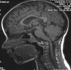
Of any of the organs in the body, the brain is the least understood, in part because it is so difficult to access and study.
As a result, relatively little is known about the underlying causes and mechanisms of psychiatric diseases, such as depression and addiction, and how best to treat them as compared with other illnesses. Fortunately, advanced imaging technologies, such as fMRI, are allowing scientists to explore the brain and its neural circuitry like never before. At the same time, new frontiers in psychiatric treatments are emerging thanks to two maturing technologies, transcranial magnetic stimulation and deep brain stimulation. These technologies allow physicians to alter the electrical activity in the brain by directly stimulating specific neural regions.
While early results are promising, much more research is needed to assess the efficacy and side effects of these technologies in patients with psychiatric illnesses. Most brain imaging is performed outside of an operating room and of limited use to neurosurgeons trying to locate areas of critical function, and current navigation methods are tedious and imprecise. As a result, some interventions, like placing probes and electrodes, are not always successful and repeat surgeries are sometimes required.
With the opening of the CSI, brain imaging can be performed during surgery, enabling surgeons to be more precise and efficient, thereby accelerating the testing and assessment of new surgical psychiatric treatments.